Squat Optimisation
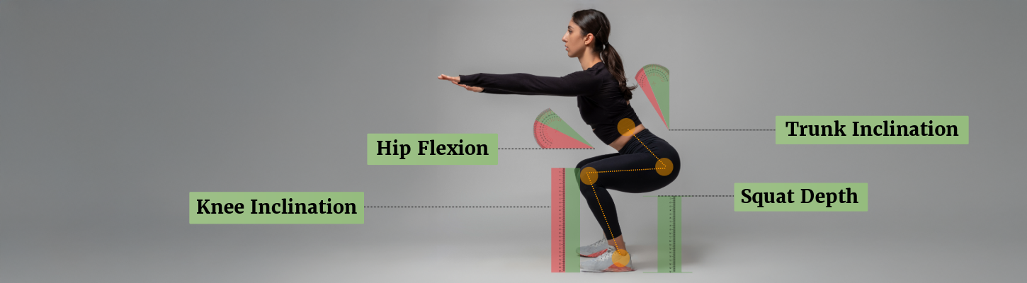

Introduction
The barbell squat is a fundamental strength exercise but also a complex, multi-joint movement that places significant mechanical demands on the spine and lower-limb joints.
Variations in technique—including trunk inclination, knee position, stance width, squat depth, and foot rotation—can substantially alter joint loading, muscle activation, and injury risk.
Evidence from motion-capture analyses, musculoskeletal modelling, and in vivo measurements shows that these variables influence compressive, shear, and rotational forces, particularly at the lumbar spine and knee joints.
This article synthesises current biomechanical research to provide evidence-based guidance for optimising squat technique while reducing unnecessary joint stress.

1. Trunk Angle
A greater forward trunk inclination, as observed during a barbell back squat, is associated with increased compressive forces on the spinal column—particularly within the lumbar region—compared to the more upright posture typically maintained in a barbell front squat.<tt>1055</tt>
Studies that quantify compressive force, shear force, and joint torque during squatting movements utilise motion capture systems in combination with musculoskeletal modelling. These models are often informed by trigonometric calculations that account for body composition and the magnitude of the external load.
Using such methods, one study estimated compressive forces at the L3/L4 spinal segment to be approximately <u>1208 N during a barbell back squat</u>, compared to <u>918 N during a barbell front squat</u>.<tt>1054</tt>
In addition, research comparing muscle activation between these two squat variations frequently reports that individuals <u>lift approximately 15–20% less absolute load during a front squat</u> than during a back squat, despite demonstrating <u>similar levels of muscle activation</u>.
A more compelling observation illustrating how trunk inclination affects the spinal column is found in a 1999 study in which a pressure sensor was surgically inserted into the L4/L5 intervertebral disc (nucleus pulposus) of a healthy adult. This allowed for direct measurement of intradiscal pressure under various postures and movements.<tt>1053</tt>
When the participant stood upright, intradiscal pressure measured 0.50 MPa; however, forward trunk inclination while standing increased this value by over 200% to 1.10 MPa.
The participant was also asked to hold a 20kg item, which increased the pressure recorded by the sensor from 1.10 MPa when held close to the body to 1.80 MPa when held 60 cm away.
This study was approved only for a single instance – for ethical reasons – but the results are compelling and support the notion that greater trunk inclination naturally places greater stress on the lumbar column.
For this reason, researchers believe a front squat, when performed with appropriate technique and loading, may reduce lumbar compressive demands compared to back squats, which may be advantageous to minimising injury risk and preserving spinal health.

2. Knee Angle
Three types of forces are considered in relation to the knee joint during a squat exercise. These are compressive force (“pressing force”), shear force (“sliding force”) and tensile force (“stretching force”).
In relation to the knee, the compressive force, or “pressing force”, is due to the femur and tibia being pressed towards each other.
The shear force, or “sliding force”, is due to the femur being displaced further anterior or posterior relative to the tibia, regulated by the anterior cruciate ligament (ACL) and posterior cruciate ligament (PCL), respectively.
The tensile force, or “stretching force”, mainly applies to the PCL, which withstands more of the tensile force, during knee flexion than the ACL.
A 2009 study found <u>no difference in shear force</u> on the knee between the front barbell squat and the back barbell squat. However, <u>compressive force was greater during a back barbell squat</u> (see figure 2).<tt>1072</tt>
Interestingly, muscle activation between both squat variations was largely the same, despite the absolute load being greater during the barbell back squat.
This paradoxical discrepancy of similar muscle activation despite a higher absolute load could be explained by a shift of "wasted forces" to joints during a barbell back squat, i.e., excess force may be applied to the joints instead of muscles.
A 2003 experimental study found torque, or “rotational force”, of the knee became greater when the knee was displaced beyond the forefeet, but torque of the hip decreased during this anterior knee displacement (see figure 3).<tt>1057</tt>
This suggests force is being increasingly shifted from the more load-tolerant hip joint to the less load-tolerant knee joint as the knee displaces beyond the forefeet – during a squat.
However, the authors emphasise that this does not necessarily mean anterior knee displacement should be discouraged. Some individuals could have unconventional anatomy (e.g., longer femurs or feet), which could necessitate the knee being displaced beyond the forefeet during a squat exercise.<tt>1061</tt>
However, the authors also state unnecessary knee displacement could place excessive forces on the posterior cruciate ligament (PCL) of the knee, increasing the risk of injury.
3. Stance
Variation in width between the feet, also known as the stance, can result in greater muscle activation in some muscles over others during a barbell squat
A 2009 experimental study defined 'stance' as a width of 0%, 150% and 200% of the greater trochanter – a bony prominence of the proximal femur.<tt>1064</tt>
The researchers measured muscle activity of the thigh and gluteus muscles during the various stance widths and found largely similar muscle activation. However, a significant <u>increase</u> in activity of the gluteus maximus was observed with a wider stance, i.e., 200% of the greater trochanter (figure 4).
A 2001 experimental study also measured the electromyographic (EMG) activity of the calf muscles during two squat exercise variations, ‘narrow’ stance and ‘wide’ stance. <tt>1063</tt>
Increased electromyographic (EMG) activity of the gastrocnemius muscle was observed during a <u>narrow stance</u> compared to a wide stance. However, there was no statistically significant difference in the activity of other muscle groups of the thigh or calf between the two stances.
The increased gastrocnemius muscle activity during a narrow stance can be explained by increased length/tension of the muscle as the knee displaces further anterior from the neutral body.
Among participants who employed a narrow stance, there was an <u>increased dorsiflexion of the ankle joint</u> (associated with greater anterior knee displacement) compared to a wider stance (see figure 5). Consequently, a narrow stance may not be optimal for those who would like to reduce forces on the knee.<tt>1074</tt><tt>1057</tt>
A 2018 study found that a <u>wider stance and a small external foot rotation</u> were associated with <u>decreased mediolateral knee displacement</u> compared to a narrow stance, which gave the chest space to incline freely instead of inclining against the thighs during the depth of the squat.<tt>1058</tt>
4. Squat Depth
The depth of squat, in research, is often used interchangeably with knee flexion and defined as the <u>angle between the thigh and shank of the lower limb</u>, with a neutral standing position being 180° and a sitting position being 90°.
Deeper squats are associated with greater muscle activation, although this is not the case for all muscle groups recruited during a squat exercise.
A 2002 study measured the electromyographic (EMG) activity in several muscles recruited during a squat – including biceps femoris, gluteus maximus, vastus medialis and vastus lateralis (see figure 6).<tt>1065</tt>
Electromyographic (EMG) readings showed the <u>activation of the gluteus maximus increased progressively as the squat became deeper</u>. These results were statistically significant, unlike the values related to the other muscles included in the electromyographic (EMG) measurements.
However, during the descent phase of a squat exercise, no muscle group was found to have higher activity despite the depth of the squat.
There is conflicting evidence as to whether a deeper squat leads to greater knee instability, for which the ligaments of the knee are mostly responsible for stabilising.
Some studies using human participants and cadavers suggest deeper squats lead to instability of the ligaments of the knee.
Such studies were not reproducible, and more recent studies suggest <u>deeper squats may actually improve knee stability</u>, at least in the long term, with improved strength and adaptation.<tt>1066</tt>
5. Foot Rotation
One of the most significant risks associated with incorrect foot rotation is the consequent effect on knee alignment, load distribution, and range of motion (RoM).
A 1% increase in knee adduction was associated with a 6.46-fold increase in risk of arthritis.
This is particularly relevant considering medial-compartment knee osteoarthritis is the most common type of arthritis in young people.<tt>1071</tt>
A 2018 study compared several foot rotations (0°, 21°, and 42°) in combination with each of the following stances: narrow stance, hip stance (normal), and wide stance during a squat exercise.
This allowed researchers to measure to what extent each rotation and stance contributed to knee misalignment and differences in load distribution. <tt>1058</tt>
The findings suggest a foot rotation of <u>0° with normal stance</u> and a foot rotation of <u>21° with wide stance</u> were the two most optimal combinations in regard to avoiding mediolateral knee deviation and achieving optimal load distribution.
The overarching recommendation was to avoid extremes, such as a narrow stance with higher rotation, which stresses the hip joints, and a wide stance with no external rotation, which stresses the knee joints.
A 2013 experimental study found no change in electromyographic (EMG) activity in the quadriceps while employing various foot rotation positions during a squat exercise.<tt>1070</tt>
Conclusion
The biomechanical demands of the barbell squat are strongly influenced by technique-related variables rather than the exercise itself.
Evidence suggests that trunk inclination, knee displacement, stance width, squat depth, and foot rotation each modify joint loading patterns and muscle activation, particularly at the lumbar spine and knee.
No single squat variation is inherently superior; rather, optimal technique depends on individual anatomy, mobility, training goals, and injury history.
By understanding how specific adjustments redistribute forces across joints, practitioners and lifters can make informed decisions to enhance performance while minimising unnecessary mechanical stress.
An evidence-based approach to squat execution is therefore essential for long-term joint health and training sustainability.